
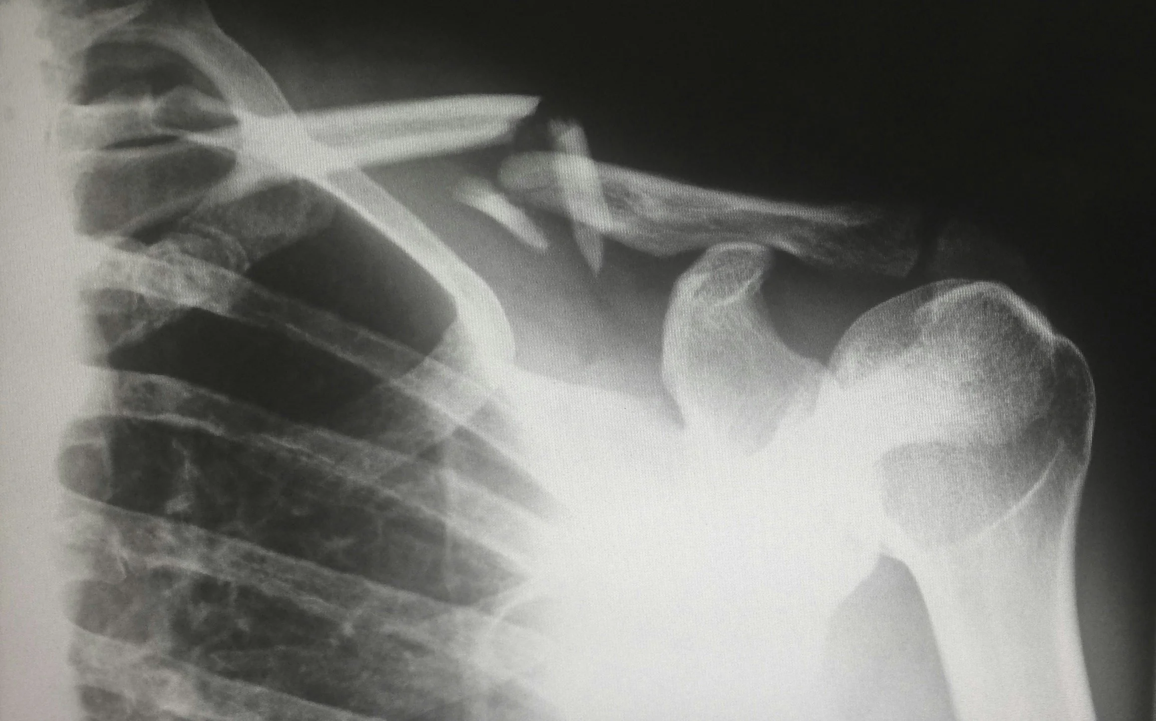
X-RAY
An X-ray is a medical imaging technique that uses a small amount of ionising radiation to create images of the inside of the body. It is commonly used to visualise bones, but it can also be used to examine other body structures such as the lungs, heart, and abdomen.
What it entails:
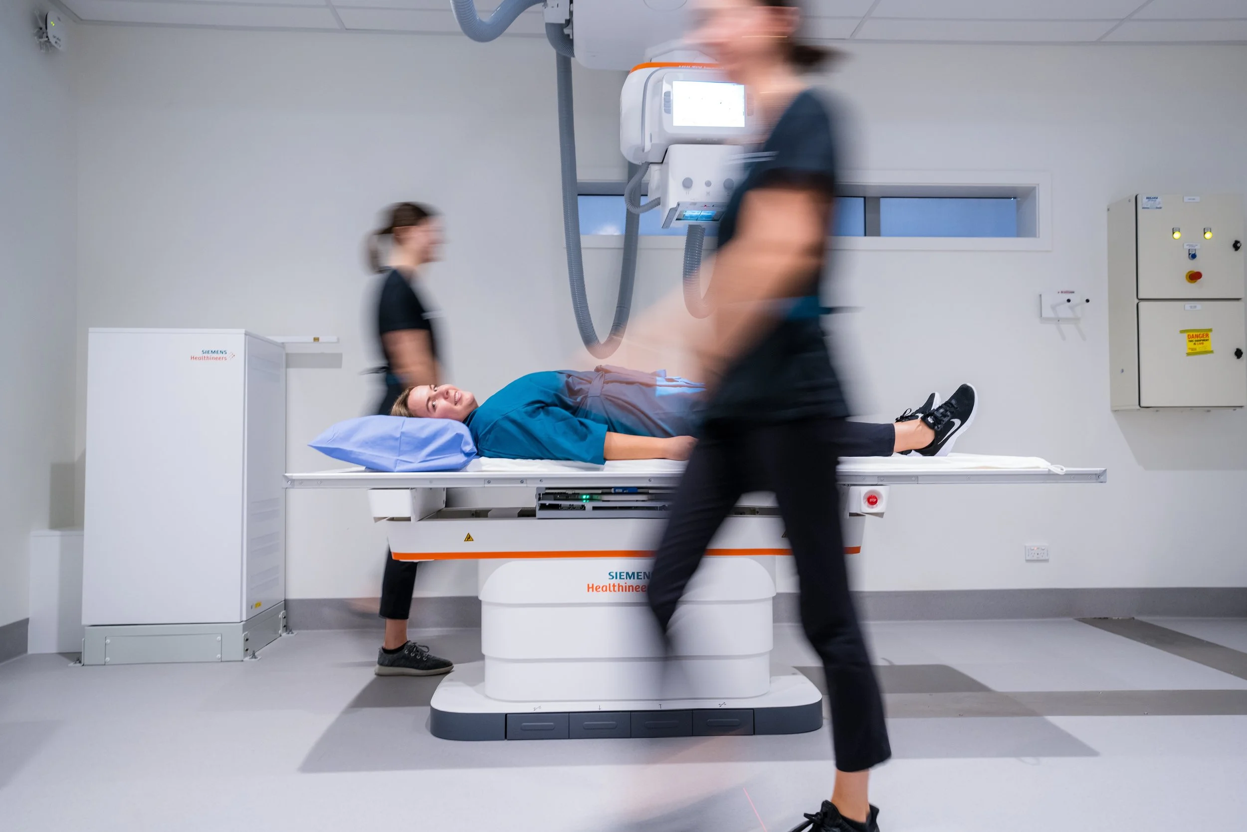
During the procedure, the patient is positioned on an X-ray table, and the X-ray machine is used to capture images by passing radiation through the body onto a detector. It is commonly used to visualise bones and other body structures, and the entire process typically takes only a few minutes. X-rays are generally safe and effective, and precautions are taken to minimise radiation exposure.
Benefits of X-ray:
Detects bone fractures and injuries: X-rays are excellent for visualising bone structures and can detect fractures, dislocations, and other skeletal injuries effectively. This makes them valuable in orthopaedic evaluations.
Diagnoses lung and chest conditions: X-rays are commonly used to identify and monitor conditions affecting the lungs and chest, such as pneumonia, lung cancer, and tuberculosis.
Quick and efficient: X-ray imaging is a relatively quick procedure, taking only a few minutes to capture images. This efficiency allows medical professionals to promptly assess a patient's condition and provide timely treatment.
Versatility: X-ray imaging can be adapted to suit various medical needs by adjusting factors like the type of X-ray used, the exposure level, and the positioning of the patient. This versatility allows for a wide range of applications.

Book an X-ray today
-
Wear comfortable clothing to your exam. You may be asked to change into a gown for the procedure.
Metal objects such as jewellery, eyeglasses, dentures or hairpins, piercings, as well metal underwires of bras or hearing aids can cause artifact on x-ray images. You may be asked by our friendly x-ray staff to remove metallic objects.
Please continue taking your medication unless instructed otherwise.
Pregnancy and breastfeeding: If you are pregnant or breastfeeding, it is important to inform our team, as special precautions may be necessary to minimise radiation exposure to the foetus or infant. Alternatively, we may delay examinations in consultation with your healthcare provider.
-
The technologist will position you on exam table or standing against the upright detector. They may use sponges or pillows to help you maintain the correct position and remain still during the exam.
Radiation exposure
X-rays involve exposure to ionising radiation, which carries a small risk of potential harm. However, the benefits of an x-ray often outweigh the potential risks, especially when it helps in diagnosing or monitoring an injury or medical condition. The amount of radiation exposure during an x-ray is typically low and varies depending on the type of exam and body part being imaged.
-
Our team of Radiologists will look at your images in detail and produce a report that will be sent out to your healthcare provider as soon as possible.