
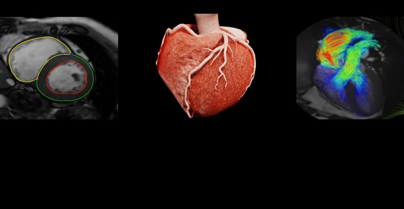
ADVANCED CARDIAC IMAGING
Cardiac imaging is a medical diagnostic technique that utilises various imaging modalities to visualise and assess the structure and function of the heart.
These modalities include techniques such as cardiac magnetic resonance imaging (cMRI), computed tomography coronary angiography (CTCA) scans, echocardiography, and nuclear imaging (myocardial perfusion). The primary objective of cardiac imaging is to provide detailed and accurate information about the heart's anatomy, blood flow, and overall performance. This enables the Cardiologist to diagnose and monitor various heart conditions, including coronary artery disease, heart valve disorders, and congenital heart defects. Early detection of heart abnormalities is vital in improving patient outcomes in the treatment and management of cardiovascular disease.
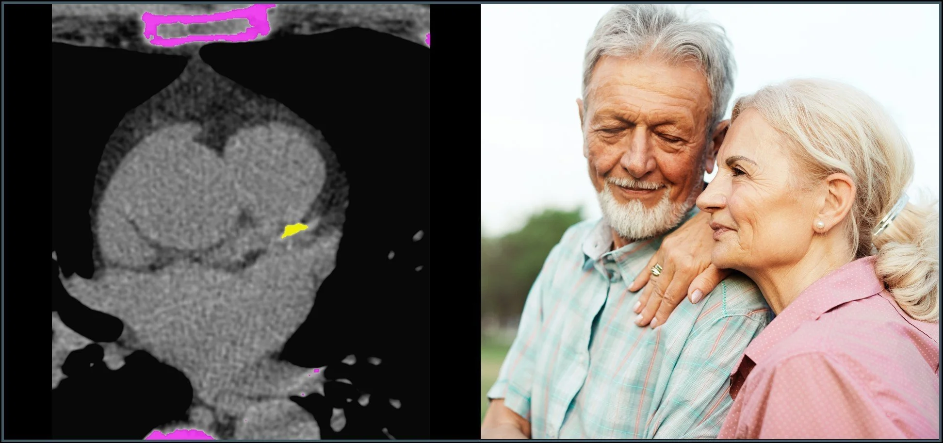
CT Coronary Arteries Calcium Score (CACS)
CT Calcium Scoring is a non-invasive imaging technique specifically designed to assess the amount of calcium deposits in the coronary arteries, aiding in the early detection of coronary artery disease (CAD). Our CT software analyses the scan data and predicts cardiovascular risk based on race/ethnicity, age and gender reference data. By quantifying calcified plaque in the coronary vessels, CT Calcium Scoring allows for a more precise evaluation of a patient's cardiac health, enabling timely preventative interventions and lifestyle modifications.
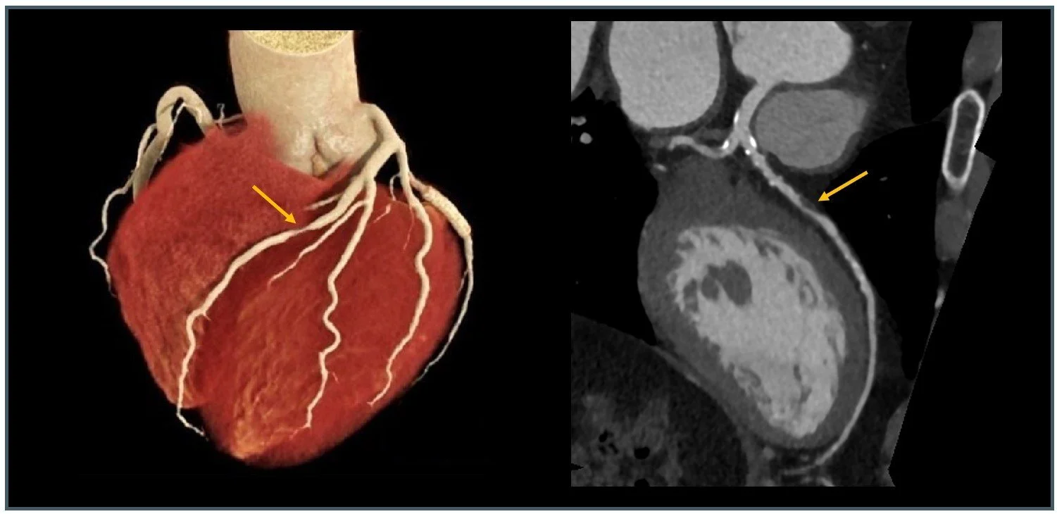
CT Coronary Angiogram (CTCA)
CT coronary angiography (CTCA) is a non-invasive medical imaging technique used to visualise the coronary arteries that supply blood to the heart muscle. Detailed three-dimensional images of the coronary arteries can be generated, assisting in the diagnosis of coronary artery disease, blockages, or other structural abnormalities. The valuable information obtained by CT coronary angiography plays a crucial role in supporting Cardiologists clinical decisions for treatment planning and patient management.
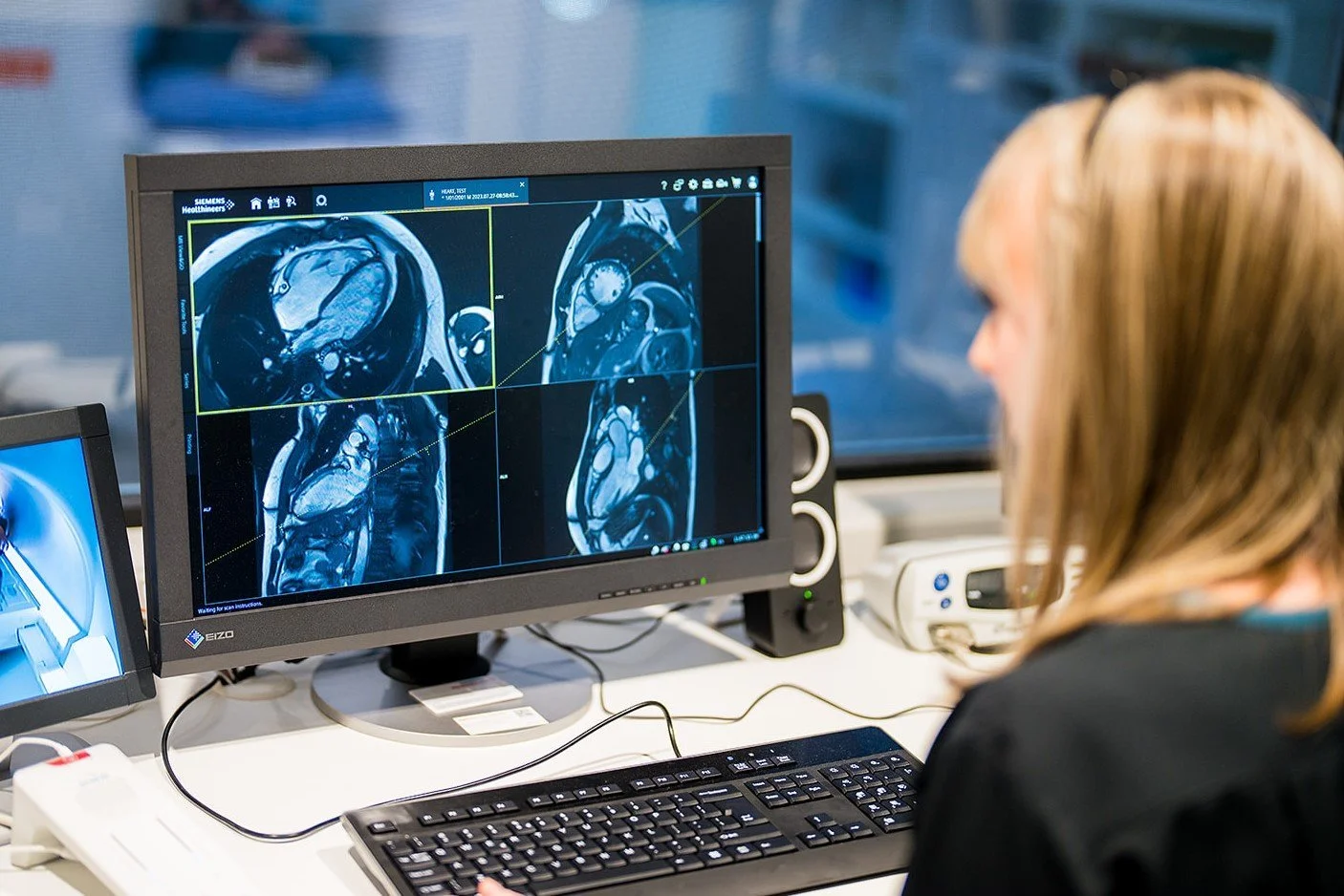
Cardiac Magnetic Resonance Imaging (cMRI)
Cardiac Magnetic Resonance Imaging (cMRI) is a non-invasive medical imaging technique that provides detailed and comprehensive visualisation of the heart's structure, function, and blood flow. It employs powerful magnets and radio waves to create highly detailed images of the heart, allowing assessment of the heart's chambers, valves, muscle tissue, and blood vessels. cMRI is particularly valuable for diagnosing various cardiovascular conditions, such as heart disease, heart attacks, and congenital heart abnormalities. It offers superior soft tissue contrast and is often used to obtain precise information about cardiac anatomy and function, aiding in the development of effective treatment plans for patients with cardiovascular disorders.
Nuclear Myocardial Perfusion Imaging
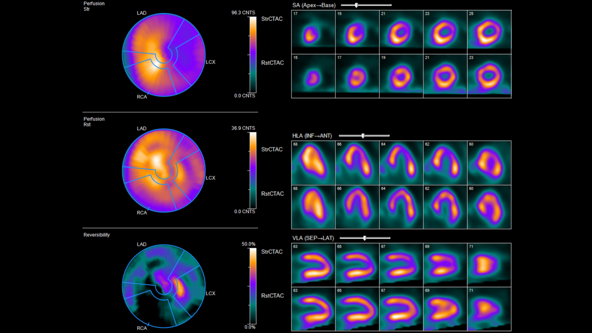
Nuclear myocardial perfusion imaging helps assess blood flow to the heart muscle and how well it is pumping. It is commonly used to investigate symptoms such as chest pain or shortness of breath, and to evaluate known or suspected coronary artery disease. This investigation is usually performed in two parts: a rest component and a stress component (either exercise or medication-induced). During each component, a small amount of injected radioactive tracer is taken up by the heart muscle, allowing it to be imaged with a specialised camera. Comparing rest and stress imaging helps nuclear medicine specialists understand how well blood reaches different areas of your heart and differentiates healthy muscle from that which is damaged or at risk of damage.

Book a Cardiac Scan today
-
Prior to the scan
Wear comfortable clothing to your exam. You will be asked to change into a gown for the procedure. Metal objects such as necklaces and metal underwires of bras may affect the CT images. You can either leave them at home or remove them before the examination.
Please continue to take all your medication unless instructed otherwise. If you have any concerns or questions about the procedure, don't hesitate to discuss them with us or your healthcare provider.
Your heart is almost constantly in motion while it beats. To get the best images we need a slow and steady heartrate.
We ask you to adhere to the following instructions prior to the appointment:
Do not drink alcohol or caffeine-based drinks (coffee, tea, energy drinks / drinks with added caffeine) and chocolate for 6 hours prior. Apart from this you can eat and drink normally, there is no need to fast.
Do not exercise before you come in on the day of your appointment.
Do not take Viagra 12 hours prior.
If you are currently taking a Betablocker, continue to take this on the day of the scan.
During the scan
When you arrive, you will be asked to change into a gown. You will lie on your back on the CT bed which will move within scanner. Your head will be outside the scanner the whole time.
Before the procedure begins, the technician will attach ECG leads to your chest. The scanner uses the ECG recording of the electrical pulses from your heart. The CT scanning is matched to the ECG, and during periods when the heart moves the least, low radiation dose images of the heart are taken, free of motion.
You will be instructed to hold your breath for each scan (approximately 15-20 seconds). It is important not to move during the scan, as it will affect the quality of the images.
After the scan
Once all the scans have been taken, the CT technologists will review the quality of the images and then you will be able to get changed and leave the department. The result of the scan will be sent to your referring physician.
-
Prior to the scan
Your heart is almost constantly in motion while it beats. To get the best images we need a slow and steady heartrate. You may be given some medication called a beta blocker to lower your heart rate. This helps to improve the image quality as there is more of a stationary period between heartbeats to obtain the data. The length of the procedure varies this is dependent on your heartrate and the need to give you betablockers prior to the scan. NB If you are given betablockers in the department these take 1 hour to work.
We ask you to adhere to the following instructions prior to the appointment:
Do not drink alcohol or caffeine-based drinks (coffee, tea, energy drinks / drinks with added caffeine) and chocolate for 6 hours prior. Apart from this you can eat and drink normally, there is no need to fast.
Do not exercise before you come in on the day of your appointment.
Do not take Viagra 12 hours prior.
Please continue to take all your usual medication.
If your Cardiologist has provided you with a Betablocker to take prior to the scan then please follow the instructions regarding this.
During the scan
When you arrive, you will be asked to change into a gown. You will lie on your back on the CT table which will move within scanner. Your head will be outside the scanner the whole time.
Before the procedure begins, the technician will attach ECG leads to your chest.
The scanner uses the ECG recording of the electrical pulses from your heart. The CT scanning is matched to the ECG, and during periods when the heart moves the least, images of the coronary arteries are taken, free of motion, so that they appear most sharp.
A desirable heart rate is vital to obtain the best quality imaging, this is why a beta blocker may be used at the time of your examination.
An intravenous (IV) line will be inserted into a vein in your arm to inject the contrast (x-ray dye) this makes your coronary arteries visible on the CT images. The contrast highlights any blockages in your coronary arteries, helping to diagnose coronary artery disease.
Just prior to the CT scan, nitroglycerin (GTN) will be sprayed under your tongue. This is used to dilate (expand) the coronary arteries to improve visualisation of the vessels.
You will be instructed to hold your breath for each scan (approximately 15-20 seconds). It is important not to move during the scan, as it will affect the quality of the images.
After the scan
Once all the scans have been taken, you will remain in the department for an additional 20 mins before the IV cannula is removed and you are allowed to leave.
Nitroglycerin occasionally causes a temporary headache; this usually resolves quickly (within 20 minutes). The result of the scan will be sent to your referring physician.
-
Prior to the scan
Clothing and accessories: If possible, leave valuables at home. You will be asked to change into a gown and remove any metallic objects, including jewellery, watches, and hairpins.
Inform the MRI technologist about any implants or devices in your body, such as pacemakers, artificial heart valves, cochlear implants, or neuro-stimulators. Some implants may be contraindicated (Not safe) for an MRI.
If a contrast agent is required for your scan, the MRI technologist will inform you about any specific instructions, such as fasting before the scan. Guidelines about eating and drinking before an MRI vary between specific examinations. Our team will let you know in advance. Take food and medications as usual unless instructed otherwise.
Please let our team know if you are or think you could be pregnant.
During the scan
The MRI technologist will complete the safety check with you. You will be asked to change into a gown and the MRI technologist will then take you into the scanner room. You will be positioned on the examination bed, and we will try to make you as comfortable as possible. ECG leads will be placed on your chest so we can view your heartbeat. An imaging device will then be placed on the chest region that will be examined. These can send and receive radio waves from your body to create the images.
The doughnut shaped MRI scanner is open at both ends. The body part being examined is placed into the center of the MRI scanner. During your examination you will be given a squeeze ball (patient safety bell) in case you need assistance during your scan. Our staff can see and hear you the whole time so please know that you are safe.
The MRI scanner makes a variety of loud noises during the scan and the MRI technologist will provide hearing protection in the form of earplugs and headphones.
Sometimes we will ask you to hold your breath during the examination. This will be important to obtain clear images of your heart.
If contrast is required for the scan, an MRI technologist or Radiologist will insert an intravenous catheter (IV line) into a small vein in your hand or arm. The contrast provides additional detailed information that helps the Radiologist or Cardiologist interpret your images.
After the scan
If you have been injected with contrast, we will ask you to stay with us for further 10 minutes to ensure your safety. We will then remove your IV line.
Please let our team know in the unlikely event that you experience any unusual symptoms during or after the examination.
Our team of Radiologists or Cardiologists will then look at your images in detail and produce a report that will be sent out to your healthcare provider as soon as possible.
-
Prior to the scan
You will be given specific instructions before your appointment, as preparation is important for accurate results. Instructions will be personalised, and typically involve fasting on the day of the test and avoiding caffeine (including coffee, tea, chocolate) for 24 hours. It may be necessary to temporarily withhold some medications.
Please provide our staff with a full list of medications, and inform us if you are pregnant, breastfeeding, have asthma or COPD, or have any allergies. Wear comfortable clothing and shoes suitable for walking if an exercise stress test is planned.
During the scan
The test usually involves two parts: imaging at rest and imaging during stress. During the stress component, you may be asked to exercise on a bicycle. If exercise is not suitable, you may receive medication that temporarily dilates blood vessels in the body, as would occur during exercise. Your heart rate, blood pressure, and ECG will be monitored throughout. You may experience mild symptoms such as warmth and flushing, shortness of breath, or headache during the stress component; these usually pass quickly.
Whilst the images are being acquired, you will lie still on a scanning bed while a camera rotates around your chest.
After the scan
You can usually return to your normal activities straight away. Drinking plenty of fluids after the test helps flush the tracer from your body. Close contact with children and pregnant women may need to be avoided for a few hours after the test; personalised advice will be provided where required.
A nuclear medicine specialist will review the images and send a report to your referring doctor, who will discuss the results with you and explain any next steps.